Understanding Brachycephalic Ocular Syndrome (BOS)
Understanding Brachycephalic Ocular Syndrome (BOS)
Dr. Louise O’Leary, MVB, DACVO.
Brachycephalic Ocular Syndrome (BOS) is a clinical condition affecting "flat-faced" dog and cat breeds (e.g., Pugs, Bulldogs, Shih Tzus, and Persian cats). Their unique skull shape results in shallow eye sockets and a flattened facial structure, leaving the eyes protruding and highly vulnerable to injury, dryness, and painful, sight-threatening diseases.
Why Does BOS Occur?
The syndrome is rooted in physical anatomy. Because the eyeballs (globes) sit so far forward, the eyelids often cannot close completely—a condition called lagophthalmos. Other contributing anatomical factors include:
● Macroblepharon & Scleral Show: Eyelid openings that are too large, exposing the "whites" of the eyes.
● Entropion & Trichiasis: Inward-rolling of the inner lower eyelid or prominent nasal skin folds that cause hair to rub directly against the cornea.
● Reduced Sensitivity & Poor Blinking: These pets often have less feeling on the eye surface and blink less frequently, meaning they may not show signs of pain even when a serious injury is present. They may sleep with their eyes open or be unable to cover their eye completely when they blink (lagophthalmos).
What Are Signs of BOS?
Symptoms can be acute or chronic and may include:
● Red Eye: The white of the eye may be red or bloodshot, often accompanied by goopy discharge.
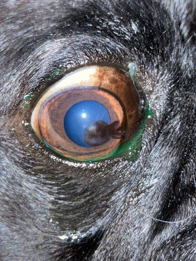
● Pigmentary Keratitis: A dark brown/black "film" that grows over the cornea due to chronic irritation in dogs, potentially leading to vision loss.
● Corneal Ulcers: Open wounds on the eye surface. In brachycephalics, these can quickly become infected, deep, or "melt," risking the loss of the eye.
● Epiphora: Chronic tearing and tear-staining caused by irritation or kinked tear ducts.
● Proptosis: An emergency where the eyeball displaces in front of the eyelids, often triggered by minimal trauma or restraint.
Diagnosis and Management
Brachycephalic pets should receive a veterinary eye exam at least once a year. Specialist exams typically involve tests to measure tear production and quality, Fluorescein Stain (checking for ulcers), and Slit-lamp Biomicroscopy for high-magnification evaluation.
Treatment Options
● Medical Management: Lifelong use of artificial tear lubricants to keep the cornea moist, and immunosuppressant drops (like Cyclosporine or tacrolimus) that reduce scarring and pigment progression, as well as improving tear quality, and reducing inflammation of the surface of the eye.
● Surgical Intervention: Medial Canthoplasty, the "gold standard" surgery. It shortens the eyelid opening at the inner corner to help the pet blink effectively, protects the globe, and stops inward-rolling lids. It improves tear film quality reducing the drying and inflammation of the surface of the eye. It can help make it easier to manage the pigment and scarring of the cornea. It can also reduce tearing. Early intervention significantly reduces the risk of future ulcers or proptosis.
Monitoring
Disease can progress rapidly in these breeds. Seek veterinary attention immediately if you notice:
● Redness, eye rubbing, or squinting (even if mild).
● Cloudiness or a blue/hazy appearance.
● Increased tearing or a change in the discharge appearance.
● A "divot" or depression on the surface of the eye.