
MY DOG IS BLIND… DOES THE EYE HAVE TO COME OUT?
Unfortunately, there are many reasons for dogs to experience blindness. Some dogs are born blind, with multiple congenital ocular abnormalities. Some experience a blinding trauma, blinding glaucoma (elevated intraocular pressure), a blinding cataract, blinding retinal disease… the list is limited only by one’s imagination.
There are 5 potential ways to treat blindness in the dog.
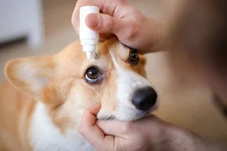
1—MEDICATIONS. If the eye is comfortable, or able to be kept comfortable with medications, a blind eye can be medicated topically and/or with medications given by mouth for as long as it remains comfortable, and the body is able to tolerate these medications. Treating with topical medications is generally ideal, as these almost always spare the body from experiencing any potential side effects.
For blind eyes that are painful, and are poorly or non-responsive to medical management, we consider the following treatment options:
2—ENUCLEATION. Enucleation involves surgical removal of the eye under general anesthesia (your dog is completely asleep). Once asleep, an injection of pain medication is administered behind the eye (called a retrobulbar block) to keep this area comfortable for several days, and to lessen the amount of anesthesia your dog must breathe in during surgery. Once the eye is removed, the orbital space (or bony cup that once held the eye) is filled with dissolving surgical foam soaked in pain medication to provide a second layer of comfort during healing. One layer of deep stitches is placed that dissolve over 2—4 weeks, followed by a second layer of stitches in the skin (these may dissolve on their own, or be removed by the surgeon once recovery is complete). Your pet should be comfortable immediately upon waking up from surgery, though there is sometimes referred tenderness or swelling for a few days in the TMJ joint (the hinge joint between the upper and lower jaw bones) on the same side, and/or just beneath where the stitches have been placed. This means that some dogs prefer to eat soft food during recovery. Recovery lasts for 10—14 days, and involves a rigid (plastic) E-collar, topical cold compresses, gentle cleaning, and medications given by mouth for comfort and to prevent infection. A biopsy is submitted to a laboratory in order to protect the healthy eye, as well as the body.

3—INTRASCLERAL PROSTHESIS PLACEMENT (ISP). ISP placement involves surgical removal of the internal contents of the eye under general anesthesia (your dog is completely asleep). Once asleep, an injection of pain medication is administered behind the eye (called a retrobulbar block) to keep this area comfortable for several days, and lessen the amount of anesthesia your dog must breathe in during surgery. The clear cornea and white outer shell of the eye are left intact, along with the eyelids and the muscles of the eyes. The only contents that are removed are the inside contents of the eye (including the lens, retina, and uveal tract – the tissues that produce glaucoma, cataract, retinal detachment, and bleeding). The surgical incision is made deep underneath the upper eyelid, and is not visible once it has been stitched closed. A small, temporary stitch in the outer corner of the upper and lower eyelids (a temporary tarsorrhaphy) is often used to protect the cornea during healing. Your pet should be comfortable immediately upon waking up from surgery, though there is sometimes referred tenderness or swelling for a few days in the TMJ joint (the hinge joint between the upper and lower jaw bones) on the same side, and/or just beneath where the stitches have been placed. This means that some dogs prefer to eat soft food during recovery. Recovery lasts for 10—14 days, and involves a rigid (plastic) E-collar, topical cold compresses, gentle cleaning, a protective topical antibiotic, and medications given by mouth for comfort and to prevent infection. A biopsy is submitted in order to protect the healthy eye. Some dogs treated with this procedure can develop dry eye (low tear production) later on in life, as they have kept the outer, natural shell of the eye, and could require tear stimulating eye drops at some point. This is a cosmetic alternative to enucleation, but cannot be performed if there is a tumor present inside of the eye, or if there is a globe perforation (hole in the cornea or the sclera) that would allow the prosthetic implant to slip out.

Right eye
4—INTRAVITREAL GENTAMICIN CILIARY BODY ABLATION (CBA). Intravitreal gentamicin involves an injection of gentamicin (an antibiotic) into the very back of the eye administered using topical numbing medication, and occasionally, a small amount of sedation. No general anesthesia or stitches are involved. This injection is intentionally toxic to the part of the eye (the ciliary body) that produces fluid inside of the eye. 60% of gentamicin CBAs are successful the first time, meaning that 40% of dogs will require re-treatment. Gentamicin must be processed by the kidneys as it leaves the body, and is highly nephrotoxic (toxic to the kidneys), meaning that some dogs can experience acute kidney injury or acute kidney failure following a gentamicin CBA, and most dogs require pre- and post-treatment intravenous fluid therapy to clear the medication from their body and protect their kidneys. 40% of dogs treated with a gentamicin CBA will develop a malignant intraocular tumor later in life with the potential to cause fatal disease elsewhere in the body. Most eyes that are successfully treated with a gentamicin CBA experience severe shrinking (phthisis bulbi), which is occasionally uncomfortable or painful, causing soreness and/or itchiness.

Right eye
5— INTRAVITREAL CIDOFOVIR CILIARY BODY ABLATION (CBA). Intravitreal cidofovir involves an injection of cidofovir (an antiviral) into the very back of the eye administered using topical numbing medication, and occasionally, a small amount of sedation. No general anesthesia or stitches are involved. This injection is intentionally toxic to the part of the eye (the ciliary body) that produces fluid inside of the eye. More than 90% of Cidofovir CBAs are successful the first time, meaning that less than 10% of dogs will require re-treatment. Cidofovir is non-toxic to the body, and requires no pre- or post- treatment intravenous fluids, with no risk to the kidneys. Cidofovir is not related to intraocular or other tumor formation following treatment. Most successfully treated eyes experience minimal to no shrinking (phthisis bulbi), and remain normally comfortable following treatment.

Right eye
Elizabeth Lutz, DVM, MS, DACVO