Signs of Age-Related Change in the Eye

Signs of Age-Related Change in the Eye
Just like human beings, animals experience many different changes within the tissues of the eye that can affect comfort and vision as aging progresses. Routine ophthalmic examinations with a veterinary ophthalmologist as part of a senior wellness program are helpful in identifying age-related changes in animals over approximately six years of age. Many age-related changes in vision can be improved with non-invasive medical treatment, once properly differentiated from more sinister diseases by a qualified veterinary ophthalmologist.
Most age-related changes within the eye become evident on ophthalmic examination in the dog around 5—6 years of age, whereas similar changes do not become evident on examination in the cat until after 10 years of age. Clinically, many dogs do not become symptomatic for these changes until after 10 years of age, whereas many cats are not symptomatic until they are teenagers.
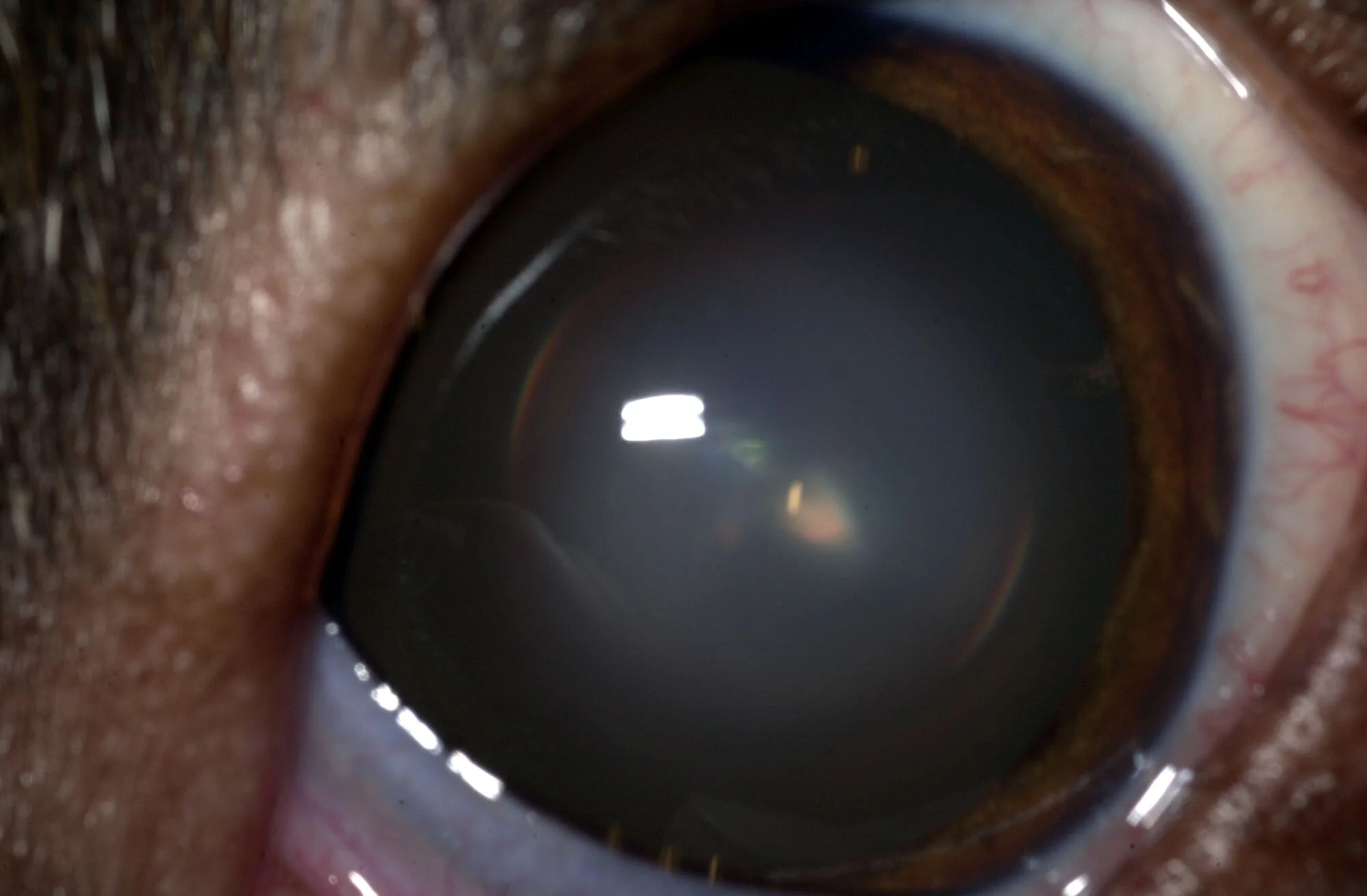
The most common age-related eye change is nuclear sclerosis (also called lenticular sclerosis). In humans, this same change is called presbyopia (and begins around 40 years of age). Nuclear sclerosis is a gradual hardening or compacting of the lens fibers with age. The fibers of the lens grow similarly to the rings of a tree, and become very dense at the center of the lens (the nucleus) with age. As this hardening occurs with age, the lens is less pliable, and becomes less able to focus. Clinically, nuclear sclerosis causes a loss in the ability to focus on objects within the near field of view. (In human medicine, this is known as farsightedness.) Some animals affected by nuclear sclerosis may bump into objects as they get very close to them. If this is happening to your pet, you should consider making an appointment with an ophthalmologist.
As nuclear sclerosis develops, the center of the lens begins to appear hazy, white, or cloudy. This is very often confused for cataract, but cannot be differentiated from a cataract without a dilated lens exam. (This means that the pupil must be dilated with specialized eye drops in order to examine the periphery or outer edges of the lens, as well as the retina.) It is critical to differentiate between nuclear sclerosis and cataract, as nuclear sclerosis is a normal, expected age-related change within the eye, and cataract is a pathologic disease change. Cataract may require surgery for vision, and almost always requires eye drops for comfort. Nuclear sclerosis does not result in pain. However, eye drops are often prescribed to help improve vision in dogs with nuclear sclerosis, and these are different from drops prescribed for cataracts.
As animals age, iris atrophy, or thinning of the muscles within the iris, also develops. The iris is the colored part of the eye. As iris atrophy develops, the inner edge of the iris (the pupil) can change from being perfectly smooth and circular to rough looking or moth-eaten. In some affected animals, the pupil will also slowly become more dilated, and less able to constrict when exposed to light. This may cause the eye to become sensitive or uncomfortable when exposed to light (photophobic). If you believe that your pet is squinting or uncomfortable in bright light, you should consider making an appointment with a veterinary ophthalmologist. It is very important to differentiate iris atrophy, which is a normal and benign age-related change, from glaucoma, which is elevated intraocular pressure inside of the eye that causes blindness, and often causes a dilated and/or misshapen pupil. Glaucoma always requires medications for the purposes of comfort and vision, and often requires surgery, whereas iris atrophy generally requires no treatment at all.
Aging humans experience a condition known as macular degeneration. The macula is a specialized structure within the retina. Domestic animals like the cat and dog do not have maculas within their retinas, and therefore, cannot experience macular degeneration. However, these animals can and do experience age-related or senile retinal atrophy that results in visual impairment. Senile retinal atrophy is characterized by thinning of the cellular layers within the retina that are responsible for vision. Initially, the retinal cells that are most responsible for night vision are affected, meaning that animals affected by senile retinal atrophy are more hesitant to navigate in dimly lit environments (at night or with the lights off), may walk more slowly, and may bump into familiar objects, but can see normally during the day or with the lights on. In some affected animals, visual impairment can progress to affect daytime and nighttime vision. If you believe that your pet is no longer seeing normally at home, you should consider scheduling an appointment with a veterinary ophthalmologist. Senile retinal atrophy must be differentiated from other diseases that are painful and can lead to permanent blindness. Fortunately, senile retinal atrophy is not painful, and visual impairment can often be lessened with oral neutraceutical supplementation that helps to slow age-related change and preserve functional vision.
Although the aging process is inevitable, there are many things that we can do to help our beloved pets age comfortably and gracefully. Routine eye examinations with a veterinary ophthalmologist are a valuable part of a wellness program for every pet at every age.