Geriatrics
Nancy M. Bromberg, VMD, MS, DACVO
Understanding Geriatrics
Several "normal" aging changes occur in the eyes of dogs and cats. Occasionally, these changes need to be differentiated from possible pathologic problems which may present in a similar manner.
LENTICULAR SCLEROSIS
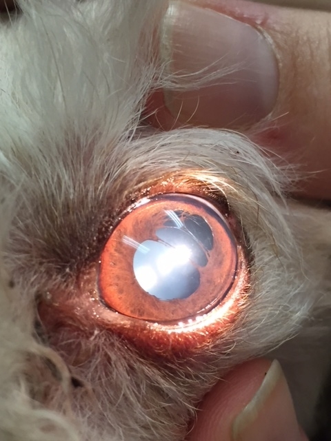
Lenticular Sclerosis and Iris Atrophy
The most common change noted in the aging animal is lenticular sclerosis, or hardening of the lens. As aging occurs in dogs, cats, and humans, a point is reached where near vision becomes a bit out of focus without the help of "reading glasses” The lack of good depth perception frequently leads to a hesitancy with stairways, decreased ability to catch toys, or an occasional nip on the finger when being given a treat. With dogs in their teens, owners may notice a "flinching" response when the animal is approached, due to decreased depth perception. Lenticular sclerosis is sometimes referred to as senile cataracts, but a cataract is a true lens opacity that obstructs vision.
IRIS ATROPHY
Thinning and atrophy of the iris sphincter muscle and/or (less frequent) the stroma of the iris is another frequent aging change. The absence of a normal pupillary light response causes an inability for the eye to control how much light is entering it, therefore occasionally causing the animal with iris sphincter atrophy to squint in bright light. Atrophy of the muscles of the pupil sphincter causes a decreased pupillary light response, and dilation of the pupil. Since the atrophy may not be symmetric in both eyes, anisocoria (different sized pupils) is common. The naturally occurring pupil sphincter atrophy needs to be differentiated from pathological abnormalities such as glaucoma, Horner's syndrome, uveitis, other cranial nerve abnormalities, and other causes of anisocoria.
ENDOTHELIAL DEGENERATION OF THE CORNEA
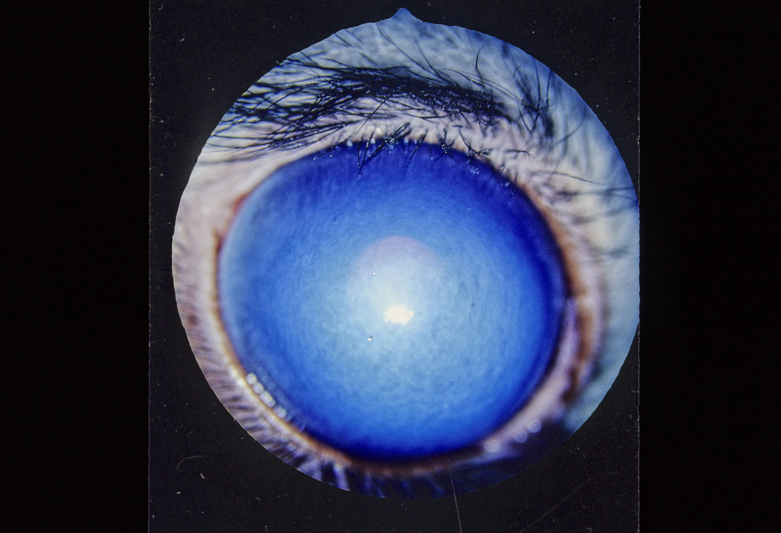
Edema due to Endothelial Degeneration
The cornea is made up of layers of collagen layers are arranged in layers, allowing light to pass through them. The inner-most layer of the cornea, the endothelium, biochemically keeps the collagen fibers dehydrated. As endothelial cells degenerate with age, the efficiency in which the remaining cells function decreases, and they allow fluid to enter the collagen. The collagen fibers swell, and the corneal becomes edematous. With the forward motion of the fluid, the fluid may accumulate between the stroma and the corneal surface epithelium, forming small bullae or "water blisters". If theses rupture, small recurrent corneal ulcerations may occur. The resulting ulcerations are slow to heal due to the edema and decreased healing due to age. This condition is known as Bullous Keratopathy. The presence of corneal edema vision is somewhat like looking through frosted glass.
RETINAL Thinning
Occasionally in the aging dog or cat, some peripheral thinning of the retina may be identified. As the photoreceptor cells of the canine and feline retina are predominantly rods, responsible for night vision, this thinning of the retina may be associated with some decrease in night vision.